Contents |
|
 Surgical Intervention in Orthodontics Problems
Introduction
Orthodontics is a growing field and as the introduction of straight wires orthodontics, a wider range of treatment is now possible. Increasingly surgical intervention is incorporated into the overall orthodontic treatment. This essay will outline the specific techniques available in Oral Surgery in the aid of orthodontics
Extraction of Teeth
Extraction of deciduous teeth – Serial extraction
The planned extraction of deciduous teeth will make sure that eruption of permanent teeth are orderly and in the right sequence
Extraction of Permanent teeth
Planned permanent teeth extraction depends on the orthodontist, whether the treatment involve extraction or non-extraction. The most common teeth extracted are premolars.
Removal of Supernumeraries
In some cases there are supernumeraries that may hinder in the successful orthodontics treatment. These teeth may be removed or auto transplanted (see later)
Surgical Removal or exposure of impacted teeth
Removal of impacted teeth
The orthodontist primarily decides removal of impacted teeth as an adjunct to the orthodontic treatment. The teeth usually removed are the impacted wisdom teeth or impacted supernumeraries. It is debatable whether wisdom teeth will influence the anterior lower crowding of the incisors.
Surgical removal of supernumeraries are however desirable if it is obstructing alignment of the other teeth in orthodontic treatment.
Special investigation may be ordered to determine the position of impacted teeth. This include
- Occlusal view
- Periapical view in Parallex- Cone in two positions relative to consecutive films in the same position. Follow the "SLOB" rule.
Exposure of impacted teeth
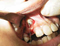
The picture above demonstrates an exposure of an impacted canine in which an othodontic button is placed and is then tracked down into the occlusion
This is relevant and is especially common to maxillary canine since it is the last tooth to erupt in that region, often it is crowded out and prevented from eruption by adjacent teeth. Surgical exposure of such teeth will give an opportunity for the orthodontist to place brackets or stainless steel loops on the impacted tooth to be drag down into its proper occlusion. However it is also common for the surgeon to create a pathway through the bone for passive eruption to occur.
It is very important, in both removal or exposure of impacted teeth, that the operator knows if the impaction is either palatal/lingually or labial/ bucally placed since burring on the wrong side could create a lot of unnecessary trauma or even a fistulation. Therefore clinical and radiographic determination of the position is utmost in importance.
It may also be important to prepare a surgical template for the palate so as to prevent edema or haematoma from occurring especially in the palate post operatively.
Surgical removal of Pathology
Often teeth are prevented from erupting to its proper position due to an underlying pathology. These may include eruption cyst, compound or complex odontomes or even ankylosis of the deciduous teeth. These have to be removed surgically before proper tooth eruption can occur
Auto-transplantation
This is a surgical procedure in which tooth from one part of the mouth is transplanted to another. Indications for such procedure are as below:
- Hypodontia – This is where there are missing tooth or teeth such as a missing central incisor, a premolar can be use as a substitute.
- Premature loss of tooth- especially true in first molar area where it may have been loss due to caries and the space is too great to be closed by the second molar. A third molar tooth is judiciously removed and a socket is prepared in the first molar area and molar is then placed and secured with 0.5 mm eyelet wire to the adjacent teeth.
However auto-transplantation is now not done as it use to due to its low success rate and largely has been superceded by placement of dental implants.
It is also important to take note that all auto-transplanted teeth need to have endodontics done, as almost always these teeth will be non-vital.
Orthognathic Surgery
This in itself a topic of its own, however it is often pointed out that orthodontist plays a big role, if not the leading role in determination and correction of the facial profile of the patient. The orthodontist determines if "masking" the result can compensate skeletal appearance by purely orthodontics or surgical interference could be of some benefit to the patient. This includes patient who has Class II or III skeletal discrepencies. It also applies to patient with maxillary excess and shows a very "gummy" smile.
Distraction Osteogenesis
A relatively new field in oral surgery, originated from orthopaedic surgery involves non-extraction orthodontics. The logic behind this is instead of removal of the premolars to compensate for lower anterior crowding, why not grow more bone to widen the jaw. The technique involves a midline mandibular osteotomy and an intra-oral distraction device placed to widen the Jaw 0.5 mm a day for the next 7 to 10 days depending on the width of the predetermined osteogenesis. The device is then left placed in its final position for the next 3 to 5 weeks for consolidation of the alveolar bone to occur and teeth will then be able to be orthodontically moved into its new position
Surgical placements of Dental Implants as Anchorage.
Recent advancements in osseointegration technology make it possible to use dental implants as anchorage points for orthodontic treatment. These implants can be use either as permanent prosthesis or removed later when the orthodontics treatment has been completed.
Dr Firdaus Hanapiah
OS/99/01
Copyright 1999, Ahmad Fariz Hanapiah![]()